- 疤痕超聲檢測儀,對(duì)疤痕規(guī)范治療意義重大,,避免了“以肉眼判斷,、憑經(jīng)驗(yàn)治療”的弊端!
- 亞細(xì)胞選擇性光熱作用原理應(yīng)用于黃褐斑治療分析
- 祛斑經(jīng)典,,穩(wěn)如磐石 雙脈寬Q開關(guān)Nd:YAG激光
- HEALITE II 嗨光丨可修復(fù)痘肌+敏感肌,,更適合術(shù)后修復(fù)~
- 讓肌膚煥然一新,,嗨光修復(fù)真的這么神奇嗎,?
- CBS云鏡皮膚檢測報(bào)告(最全解析)
- 王展博士介紹全新一代SPECTRA2020
- 這7種類型的疤痕,,用點(diǎn)陣激光去除效果真好!
- 點(diǎn)陣激光祛疤效果怎么樣?會(huì)復(fù)發(fā)嗎,?
- 疤痕超聲檢測儀,對(duì)疤痕規(guī)范治療意義重大,,避免了“以肉眼判斷,、憑經(jīng)驗(yàn)治療”的弊端!
- 亞細(xì)胞選擇性光熱作用原理應(yīng)用于黃褐斑治療分析
- 祛斑經(jīng)典,,穩(wěn)如磐石 雙脈寬Q開關(guān)Nd:YAG激光
- 能修復(fù)痘肌,、敏感肌,號(hào)稱“膠原光”的嗨光來啦!
- 【皮膚美容】1064nm激光聯(lián)合醫(yī)用敷料治療面部皮膚光老化療效研究
- CBS云鏡皮膚檢測報(bào)告(最全解析)
- 王展博士介紹全新一代SPECTRA2020
- 這7種類型的疤痕,用點(diǎn)陣激光去除效果真好,!
- 點(diǎn)陣激光祛疤效果怎么樣,?會(huì)復(fù)發(fā)嗎,?
20 MHz高頻超聲在瘢痕評(píng)估中的應(yīng)用
本文來源:《中華整形外科雜志》2023年6月 第39卷 第6期
DOI:10. 3760 / cma.j.cn114453-20220320-00075
作者單位:空軍軍醫(yī)大學(xué)第一附屬醫(yī)院燒傷與皮膚外科, 西安710032
通信作者:李娜,,Email:linaxjss@163.com
【摘要】
目的 探討20 MHz高頻超聲在瘢痕厚度及形態(tài)學(xué)評(píng)估中的作用,。
方法 對(duì)2019年4月至2020年12月空軍軍醫(yī)大學(xué)第一附屬醫(yī)院燒傷與皮膚外科診治的燒創(chuàng)傷后瘢痕形成初期(<1個(gè)月)、增生期(1~6個(gè)月)和消退期(>6個(gè)月)患者的臨床資料進(jìn)行回顧性分析,。全部納入患者均進(jìn)行了20 MHz高頻超聲,、溫哥華瘢痕量表(VSS)及病理組織學(xué)3種方式的評(píng)估。超聲檢查時(shí)在瘢痕處選擇3個(gè)點(diǎn)進(jìn)行瘢痕厚度測量,,記錄平均值,;在超聲測量部位的瘢痕組織取標(biāo)本,進(jìn)行HE染色和Masson染色,,觀察并測量瘢痕厚度;由2名醫(yī)生采用VSS對(duì)瘢痕的厚度進(jìn)行評(píng)估,,記錄平均值,。分別比較瘢痕形成初期、增生期,、消退期采用上述3種方式測評(píng)的瘢痕厚度的差異,;同時(shí),對(duì)比高頻超聲聲像圖特征與病理組織形態(tài)學(xué)的關(guān)系,。正態(tài)分布計(jì)量資料以±s表示,,3組間比較采用單因素方差分析,組間兩兩比較采用SNK-q檢驗(yàn),;計(jì)數(shù)資料采用卡方檢驗(yàn)進(jìn)行分析,。
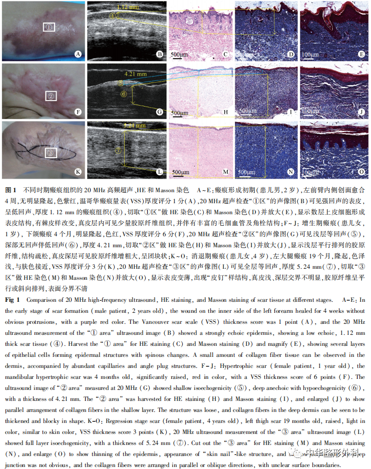
結(jié)果 共納入224例患者,男91例,, 女133例,,年齡1~34歲,平均25.7歲,;瘢痕形成初期患者79例,、增生期102例,、 消退期43例。(1)在瘢痕形成初期,,20 MHz超聲測量的瘢痕厚度為(2.01±0.68)mm,,VSS評(píng)估厚度為(1.72±0.49)mm,病理測量厚度為(2.11±0.45)mm,;在增生期瘢痕,,20 MHz超聲測量厚度為(4.11±0.73)mm,VSS評(píng)估厚度為(3.02±0.47)mm,,病理測量厚度為(4.27±0.44)mm,;在消退期瘢痕,20 MHz超聲測量厚度為(1.74±0.64)mm,,VSS評(píng)估厚度為(1.77±0.61)mm,,病理測量厚度為(1.71±0.67)mm。對(duì)于3個(gè)時(shí)期的瘢痕,,20 MHz高頻超聲測量的瘢痕厚度與病理測量厚度的差異均無統(tǒng)計(jì)學(xué)意義(P均>0.05),;而在瘢痕形成初期及增生期,VSS評(píng)估的厚度值與20 MHz高頻超聲,、病理測量厚度的差異均有統(tǒng)計(jì)學(xué)意義(P均<0.05),。(2)在瘢痕形成初期,高頻超聲顯示表皮厚度與正常表皮接近且呈高亮強(qiáng)回聲,,但表皮與真皮層之間有約<1 mm厚的條形低回聲或無回聲區(qū),,形似真皮水腫;病理組織學(xué)顯示,,該期瘢痕的表皮有棘皮樣改變,,真皮層內(nèi)有豐富的毛細(xì)血管,并有少量膠原纖維組織,。在增生期瘢痕,,表皮仍呈強(qiáng)回聲,真皮層內(nèi)呈不均勻回聲,,真皮淺層呈明顯等回聲,,而深層表現(xiàn)無回聲或低回聲;病理顯示,,表皮薄而光滑,,角化明顯,真皮淺層可見與表面平行,、排列規(guī)則的膠原纖維,,真皮深層可見膠原纖維增多增厚,呈結(jié)節(jié)狀,、漩渦狀,。在消退期瘢痕,,表皮呈強(qiáng)回聲,真皮層與皮下組織之間無明顯分界,,呈均勻等回聲,;病理顯示,表皮變薄,,出現(xiàn)“皮釘”樣結(jié)構(gòu),,真皮淺、深層交界不明顯,,膠原纖維呈平行或斜向排列,,表面分界不清。
結(jié)論 20 MHz高頻超聲對(duì)瘢痕厚度的評(píng)估較VSS更準(zhǔn)確,,且可反映瘢痕內(nèi)膠原分布,、水分比例情況,相較于病理檢查具有無創(chuàng),、快捷的優(yōu)點(diǎn),,是評(píng)估瘢痕厚度及形態(tài)學(xué)的有效手段。
【關(guān)鍵詞】瘢痕,;超聲檢查,;高頻超聲;病理學(xué),;Masson染色,;溫哥華瘢痕量表;瘢痕厚度
Application of 20 MHz high-frequency ultrasound in scar evaluation
Bai Lu, Shi Xueqin, Yang Li, Zhao Wenli, Li Na, Han Juntao, Hu Dahai
Department of Burn and Cutaneous Surgery, the First Affiliated Hospital of Air Force Medical University, Xi’an 710032, China
Corresponding author: Li Na, Email: linaxjss@163.com
【Abstract】
Objective To investigate the role of 20 MHz high-frequency ultrasound in evaluating scar thickness and morphology.
Methods The clinical data of patients with the initial stage of scar formation after burn trauma (<1 month), hypertrophic scar (1-6 months) and atrophic scar (>6 months) treated by the Department of Burn and Cutaneous Surgery, the First Affiliated Hospital of Air Force Medical University from April 2019 to December 2020, were retrospectively analyzed. All patients were evaluated by 20 MHz high-frequency ultrasound, histopathology and Vancouver scar scale (VSS). Three measurement points were randomly selected at the scar during ultrasonic examination, and the average value was recorded as the ultrasonic thickness measurement value. The scar tissue samples were collected from the site of ultrasonic examination, and HE staining and Masson staining were performed. At the same time,scar thickness was evaluated by two physicians using VSS. The difference of scar thickness assessment result among the 3 method in patients at the initial stage of scar formation, hypertrophic scar and atrophic scar was compared. Meanwhile, the relationship between the characteristics of 20MHz high-frequency ultrasound and histopathology was compared. The measurement data of normal distribution were expressed as Mean±SD. One-way ANOVA was used for comparison among three groups, and SNK-q test was used for pairwise comparison between groups. Counting data were analyzed by Chi-square test.
Results A total of 224 patients were included, including 91 males and 133 females, aged from 1 to 34 years, with an average age of 25.7 years. There were 79 patients at the initial stage of scar formation, 102 at the hypertrophic stage, and 43 at the atrophic stage. (1) In the initial stage of scar formation, the thickness measured by 20 MHz ultrasound was about (2.01±0.68) mm, the thickness evaluated by VSS was (1.72±0.49) mm, and the thickness measured by pathological section was (2.11±0.45) mm. In the hyperplastic scar stage, the thickness measured by 20 MHz ultrasound was (4.11±0.73) mm, the thickness evaluated by VSS was (3.02±0.47) mm, and the thickness measured by pathological section was (4.27±0.44) mm. In the atrophic scar stage, the thickness measured by 20 MHz ultrasound was (1.74±0.64) mm, the thickness measured by VSS was (1.77±0.61) mm, and the thickness measured by pathological section was (1.71±0.67) mm. For scars in the above three periods, there was no statistical significance between scar thickness measured by 20 MHz high-frequency ultrasound and that measured by pathological sections(all P<0.05). In the initial stage of scar formation and hypertrophic stage, the thickness evaluated by VSS was significantly different from that measured by 20 MHz high-frequency ultrasound and pathology (all P<0.05), respectively. (2) Echo intensity was evaluated by ultrasound. In the initial stage of scar formation, the thickness of the epidermis shown by high-frequency ultrasound was close to that of the normal epidermis and presented a high-intensity echo, but there was a strip of echoless or no echo zone of <1mm between the high-intensity echo epidermis and dermis, which looked like dermal edema. Pathology showed that there were acanthoid changes in the epidermis of the scar at this stage, rich capillaries and a small amount of collagen fibrous tissue in the dermis. In the hyperplastic scar stage, the scar epidermis still showed strong echo, while the dermis showed uneven echo, the superficial dermis showed obvious isoecho, and the deep dermis showed no echo or hypoecho. Pathology showed that the epidermis was thin and smooth, and keratosis was obvious. Collagen fibers parallel to the epidermis could be seen in the superficial layer of the dermis, with regular arrangement. Collagen fibers were increased and thickened in the deep layer of the dermis, in the shape of nodules and swirls. In the atrophic scar stage, the scar epidermis presented a strong echo, and there was no obvious demarcation between the dermis and subcutaneous tissue, presenting a uniform echo. Pathological findings showed that the epidermis became thinner with a “skin nails”-like structure, the junction between the superficial and deep dermis was not obvious, and the collagen fibers were arranged in parallel or oblique direction, and the surface boundary was unclear.
Conclusion 20MHz high-frequency ultrasound is more accurate than VSS in the assessment of thickness of hypertrophic scar, and can reflect the collagen content and moisture ratio in scar. Compared with pathological examination, it has the advantages of non-invasive and fast, and is an effective means to evaluate scar thickness and morphology.
【Key words】Cicatrix; Ultrasonography; High-frequency ultrasound; Pathology; Masson staining; Vancouver scar scale; Scar thickness
Disclosure of Conflicts of Interest: The authors have no financial interest to declare in relation to the content of this article.
Ethical Approval: This study was conducted in accordance with the Helsinki Declaration.
瘢痕是燒創(chuàng)傷后組織修復(fù)的必然結(jié)果,。近年來瘢痕防治越來越受到關(guān)注,,治療方法也逐漸增多。在常規(guī)的硅酮藥物,、壓力治療的基礎(chǔ)上,,激光療法,、肉毒毒素或糖皮質(zhì)激素局部注射等也獲得了該領(lǐng)域?qū)<业恼J(rèn)可,,并寫入瘢痕治療的指南中[1-2]。但個(gè)體差異,、醫(yī)生的治療經(jīng)驗(yàn),、治療方案的選擇等會(huì)導(dǎo)致瘢痕治療效果參差不齊。準(zhǔn)確,、有效的瘢痕評(píng)估是治療方案選擇,、技術(shù)參數(shù)改進(jìn)、瘢痕量化管理不可或缺的重要組成部分,,因此提高瘢痕評(píng)估的準(zhǔn)確度顯得尤為重要,。溫哥華瘢痕量表(Vancouver scar scale, VSS)是瘢痕評(píng)估簡單,、快捷且常用的方法[3],但對(duì)于瘢痕增生初期的水腫,、未突出體表的皮下瘢痕,,其厚度不易判斷,很難準(zhǔn)確評(píng)估,;且在瘢痕的質(zhì)地評(píng)分上,,“柔軟”與“柔順”的區(qū)別也很難把握。B型超聲是評(píng)估組織厚度,、組織結(jié)構(gòu)最常用且準(zhǔn)確快捷的方法,。高頻皮膚超聲因兼顧探測深度、表皮-真皮界限而廣泛用于臨床皮膚檢測[4],。瘢痕組織主要分布在近似真皮層及淺筋膜層,,20MHz高頻超聲是觀察表皮、真皮及淺筋膜層皮損的最佳監(jiān)測設(shè)備[5-6],。本研究通過對(duì)燒創(chuàng)傷后瘢痕形成初期,、增生期和消退期患者的20MHz高頻超聲、病理組織學(xué),、VSS評(píng)估情況進(jìn)行分析比較,,探討20MHz高頻超聲作為瘢痕常規(guī)評(píng)估工具的可行性。
資料與方法
一,、 資料選擇
收集2019年4月至2020年12月空軍軍醫(yī)大學(xué)第一附屬醫(yī)院燒傷與皮膚外科診治的燒創(chuàng)傷后瘢痕形成初期(<1個(gè)月),、增生期(1~6個(gè)月)和消退期(>6個(gè)月)患者的臨床資料,進(jìn)行回顧性分析,。納入標(biāo)準(zhǔn):瘢痕形成初期,、增生期、消退期患者,,無明顯瘢痕攣縮畸形及功能障礙,。排除標(biāo)準(zhǔn):近3個(gè)月使用糖皮質(zhì)激素類藥物;測量部位伴有病毒或細(xì)菌感染,;懷孕或哺乳,;不能按期隨訪。
患者或家屬對(duì)研究知情,,并同意將資料用于本研究,。本研究已參考赫爾辛基宣言。
二,、 方法
分別比較瘢痕形成初期,、增生期、消退期患者采用20MHz高頻超聲、病理組織學(xué)及VSS 3種方式評(píng)估的瘢痕厚度的差異,;同時(shí),,對(duì)比20MHz高頻超聲聲像圖特征與病理組織形態(tài)學(xué)的關(guān)系。
(一)20MHz高頻超聲
所有患者瘢痕部位檢測前與患者進(jìn)行溝通,,介紹超聲評(píng)估的目的,,檢測前清潔瘢痕皮膚,使用單反數(shù)碼相機(jī)(佳能EOS 650D型)拍照并存檔,。所有病變均采用高頻超聲皮膚診斷系統(tǒng)(天津邁達(dá)醫(yī)學(xué)科技股份有限公司,,型號(hào)MD-3000S)進(jìn)行檢查。如果瘢痕是不規(guī)則的,,以瘢痕最厚區(qū)域代表該部位瘢痕的厚度,,檢查中用20 MHz線性探頭選擇3個(gè)測量點(diǎn),測量瘢痕厚度和回聲強(qiáng)度,,記錄厚度平均值,。通過與周圍正常組織的比較來評(píng)估瘢痕的回聲強(qiáng)度,分為4種:高回聲,、等回聲,、低回聲及混合回聲。
(二)VSS評(píng)估
所有瘢痕由2名醫(yī)生采用VSS進(jìn)行評(píng)分,,以平均值作為該瘢痕的評(píng)分值,。主要從色澤、厚度,、血管分布,、柔軟度4個(gè)指標(biāo)對(duì)瘢痕進(jìn)行評(píng)估,量表總分為15分,,評(píng)分越高表示瘢痕越嚴(yán)重,。色澤:0分為正常顏色,1分為淺白色或淺粉紅色,,2分為深淺混集,,3分為深色;厚度:0分為正常,,1分為>0~1 mm,,2分為>1~2 mm,3分為>2~4 mm,,4分為>4 mm,;血管分布:0分為正常,,1分為粉紅色,,2分為紅色,3分為紫色,;柔軟度:0分為正常,,1分為柔軟,,2分為少許拉緊,3分為質(zhì)硬,,4分為牽拉關(guān)節(jié)彎曲,、關(guān)節(jié)很難伸直,5分為形成永久性軟組織攣縮如關(guān)節(jié)畸形,。
(三)瘢痕組織病理染色
在超聲測量部位的3個(gè)點(diǎn)中選擇最接近平均值的點(diǎn),,切除瘢痕組織取標(biāo)本。獲取瘢痕組織后縱行剖開,,以4%多聚甲醛固定并編號(hào),,石蠟包埋,厚度為5 μm進(jìn)行連續(xù)切片,,然后進(jìn)行脫蠟,、透明、HE及Masson染色,,光鏡下觀察并測量瘢痕厚度,。
三、統(tǒng)計(jì)學(xué)處理
采用SPSS 18.0統(tǒng)計(jì)軟件進(jìn)行數(shù)據(jù)分析,?;颊吣挲g、瘢痕厚度為正態(tài)分布計(jì)量資料,,以±s表示,,3組間比較采用單因素方差分析,采用SNK-q檢驗(yàn)進(jìn)行組間兩兩比較,;患者性別構(gòu)成比,、皮膚類型構(gòu)成比為計(jì)數(shù)資料,以例(%)表示,,采用卡方檢驗(yàn)進(jìn)行分析,。以P<0.05為差異有統(tǒng)計(jì)學(xué)意義。
結(jié) 果
一,、一般資料
......


